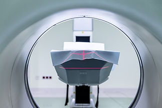
MRI
Breast MRI uses magnetic fields and contrast agents to provide highly detailed images of breast tissue. It is especially valuable for screening high-risk patients and clarifying findings from other imaging modalities. Below, you’ll find information on indications, protocols, and interpretation to guide clinical decisions.
Molecular Resonance Imaging (MRI)
What is it?
Magnetic Resonance Imaging (MRI) utilizes strong magnetic fields and radiofrequency waves to generate highly detailed, cross-sectional images of breast tissue.
How Does it Work?
MRI typically involves an intravenous (IV) injection of a contrast agent (gadolinium), which highlights blood vessels and areas of increased or abnormal blood flow, including leaky vessels that may indicate malignancy.
Best for:
Women at elevated breast cancer risk, particularly for interval cancer detection and/or for women with dense breasts.
Clinical Performance:
High Sensitivity: MRI demonstrates sensitivity between 71−100%, though specificity varies from 81−98% depending on patient population and protocol. (1)
-
Abbreviated MRI (AB MRI): Comparable sensitivity to full-protocol MRI with reduced cost and scan time. (2)
-
BRAID Study: AB MRI detected three times more invasive cancers than Automated Breast Ultrasound (ABUS), with detected tumors being approximately half the size. Detection rates were similar to contrast-enhanced mammography (CEM). (3)
Limitations:
-
Higher cost compared to other imaging modalities (though AB MRI is more cost-effective).
-
Requires contrast dye, which some patients may not tolerate or be amenable towards.
-
May result in a higher rate of false positives, leading to additional diagnostic testing.
Regulatory Status:
-
Full-protocol MRI is approved and recommended annually in addition to a mammogram for women at high risk for breast cancer.
-
Abbreviated MRI is gaining traction, but standard protocols are still under development.

